Contact Us to SCHEDULE A DEMO

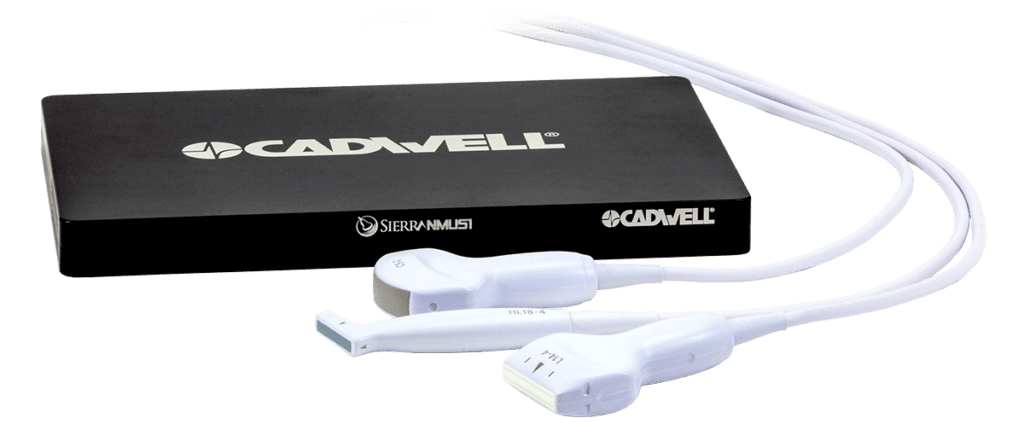
Sierra NMUS1 Ultrasound
Complement your electrodiagnostic testing with Sierra NMUS1 Ultrasound
Sierra NMUS1™ is an advanced ultrasound imaging tool designed specifically for neuromuscular diagnostics, musculoskeletal examinations, and augmented needle guidance. Sierra NMUS1 and the NMUS+ protocol in Sierra® software provide innovative and sophisticated algorithms and dedicated imaging and diagnostic features.
Sierra NMUS1 is completely integrated into the Sierra software NMUS+ protocol for fast and accurate diagnoses, enabling high image quality, an intuitive protocol-specific user-interface, a simple integrated workflow and results, and muscle quantification mode.
With Sierra NMUS1, you can:
- Instantly transition from NCS or EMG testing to imaging.
- Utilize combined protocols with improved image quality, integrated measurements, and tissue quantification to increase diagnostic accuracy and reduce examination time.
- Combine all results into a single report to clearly communicate findings.
Sierra NMUS1 is a powerful option for point-of-care imaging during neurodiagnostic examinations.
Sierra NMUS1 Ultrasound
Sierra NMUS1 is a high-performance ultrasound with variable transmit frequencies (2-18 MHz) to deliver advanced image quality and resolution. It offers eight simultaneous beamformers for fast imaging in B-Mode and four simultaneous beamformers in Color Flow Doppler Mode.
Image Enhancement & Processing Capabilities
- Spatial Compounding
- Edge Enhancement
- Basic Speckle Reduction
- Advanced Speckle Reduction
- Harmonic Scanning
- Trapezoid View
- Algo8 Clarity Enhancement
Display/Operating Modes
- B-Mode
- Color Flow Doppler
- M Mode
- Q Mode
- PW Doppler Mode
Additional Features
- Image Magnification: Zoom from 1:1 to 30:1 (real-time and frozen)
- Cine Review: 10 seconds
- Focus Position: 16 levels to adjust
- Focus Number: Max 4
Ultrasound Probes for Sierra NMUS1
Three interchangeable handheld probes (transducers) are available for use with Sierra NMUS1.
- Linear Ultrasound Probes are used to create high resolution images of superficial structures near the body surface, like vascular imaging, and are also called vascular probes. Linear probes are often used to measure movement in a linear fashion, i.e. scanning along a nerve. Different linear transducers are able to measure different movements.
- Linear Ultrasound (Vascular) Probes are used to create high-resolution images of superficial structures near the body surface.
- Curvilinear Ultrasound Probes: Curvilinear – or convex – ultrasound probes use lower frequencies for deeper penetration and wider depth of field for viewing larger muscles and deeper structures.
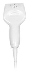
L14-4 Linear Probe
- Frequency Standard Range:
4-14 MHz, Max: 16 MHz, Central: 9 MHz - Elements:
192 Piezo Electric, 0.2 mm Pitch - Aperture: 38.4 mm
This commonly used probe comes standard with the Sierra NMUS1.
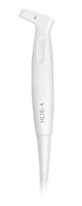
HL18-4 Linear Hockey Stick Probe
- Frequency Standard Range:
4-18 MHz, Max: 20 MHz, Central: 10 MHz - Elements:
128 Piezo Electric, 0.2 mm Pitch - Aperture: 25.6 mm
This broad frequency linear probe offers high sensitivity and deep penetration for scanning superficial, deep, and difficult to access structures.
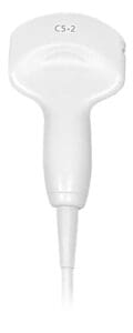
C5-2 Curvilinear Probe
- Frequency Standard Range:
2-5 MHz, Max: 6 MHz, Central: 3 MHz - Elements:
160 Piezo Electric, 0.38 mm Pitch - Field of View: 70 degrees
What does integrated ultrasound mean?
Concurrent Use
Hear, see and stimulate for more accurate injections and needle guidance.
Combine imaging with any nerve study and create a new and unique protocol.
Integrated Settings
Pre-define structures with specific image presets.
Save time with labeling.
Improve clarity of specific structures.
Integrated Controls
Adjust image settings.
Record and review snapshots and videos.
Switch between B, M, Doppler and Comparisons modes.
Integrated Workflow
Switch between US, EMG and NCS with the push of a button.
Include imaging sites in your NCS and EMG studies.
Include US images and measurements in your EDX report.
Cadwell Whitepaper: The Case for Neuromuscular Ultrasound
Recent developments in high-frequency ultrasound imaging technology have resulted in neuromuscular ultrasound (NMUS) becoming another important component of neurodiagnostic evaluations. Ultrasound can help diagnose common entrapment neuropathies and localize other nerve problems. Combined with EMG or NCS, you can increase your diagnostic certainty quickly, safely, and without patient discomfort.
Download materials, get software updates, and request hardware and software features
Shop for all the EMG electrodes and accessories you need
Explore Cadwell EDX products
The Sierra® family of products are the ideal solution for Chemodenervation, EMG, EPs, NCS, Ultrasound, and Electrodiagnostics in Urology.
Request an EMG, NCS, EP, and Ultrasound Demonstration
Product availability may vary between different countries and markets.
Please contact Cadwell for additional information.

